Microscopic analysis of patient samples is a recommended method by the World Health Organization (WHO) for diagnosing diseases and providing appropriate treatment. However, the lack of skilled microscopists in endemic regions leads to issues such as heavy workload and visual impairments.
Mr. Frank Acquah, a senior medical laboratory technician at the University of Cape Coast UCC Hospital, highlighted several causes of inaccurate diagnoses, including inexperience, lack of necessary tools, improper sampling, and workload. To overcome these challenges, seeking a second opinion in cases of uncertainty is advised.
Accurate diagnosis is crucial in providing proper care to patients as it helps identify conditions that may not be visible to the naked eye. Dr. Simon Osei-Frimpong, a family health physician, emphasized the risks associated with wrong diagnoses, including prescribing incorrect medications, which can lead to prolonged recovery, resistance, or even death. Inaccurate diagnoses are particularly prevalent in the African Sub Region, where tools and innovations for diagnosing Neglected Tropical Diseases are scarce, especially in rural areas.
Schistosomiasis and soil-transmitted helminth (STH) infections are part of the Neglected Tropical Diseases and affect millions of people worldwide, primarily those living in impoverished communities with limited access to clean water, sanitation, and hygiene.
Sub-Saharan Africa carries the highest burden of these diseases, with approximately 252 million people affected by schistosomiasis and an estimated 1.5 billion people (24% of the global population) infected with STH.
Schistosomiasis is typically diagnosed by detecting parasite eggs in stool or urine samples, while blood or urine samples indicate infection through the presence of antibodies or antigens. However, obtaining accurate diagnoses in rural communities is challenging due to the lack of necessary tools in primary healthcare facilities.
In response to these challenges, researchers at Delft University of Technology have developed an innovative tool called the schistoscope.
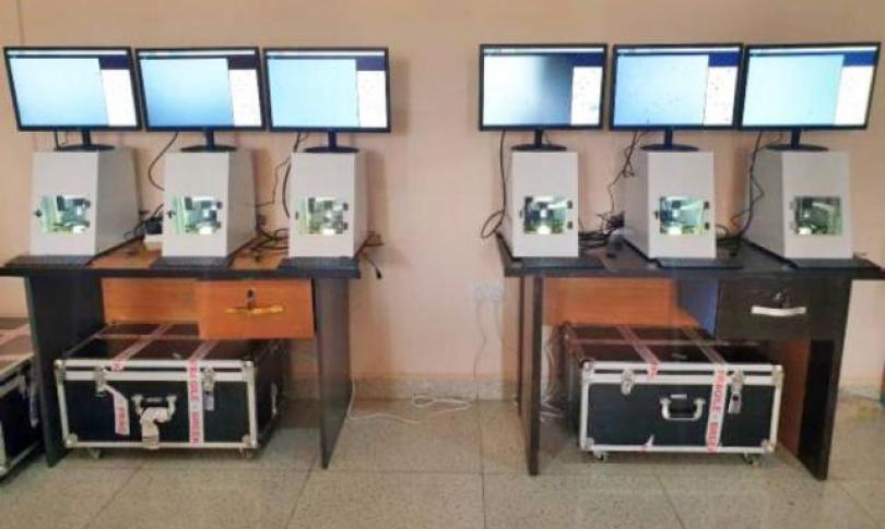
Powered by artificial intelligence, this digital microscope promises to revolutionize the diagnosis and monitoring of schistosomiasis and STH infections. The schistoscope automates the microscope process and utilizes AI for sample analysis, capturing and analyzing microscopic images of parasite eggs to provide accurate diagnoses while reducing the burden on microscopists, Dr Prosper Oyibo told Science Journalism Ghana.
It also enables disease mapping, surveillance, and determination of disease interruption, he added.
The schistoscope functions by preparing patient samples on a glass slide using urine filtration and fecal smear techniques for urogenital schistosomiasis and intestinal helminth infections, respectively. The prepared sample is then inserted into the device, and Field of View (FoV) images are captured using a digital camera, Oyibo explains.
An embedded computer equipped with AI algorithms processes the images, accurately detecting and counting parasite eggs in the sample. The diagnosis status (positive or negative) and infection intensity (eggs per mL and eggs per gram) are displayed on a screen.
Currently undergoing validation and testing in collaboration with research partners in Nigeria and Gabon, the schistoscope aims to be accessible in primary healthcare centers in endemic regions, providing essential diagnostic capabilities to those in need.
Overcoming implementation challenges, such as local manufacturability and maintenance, the researchers are collaborating with local universities and technology makerspaces to enable co-creation and develop a design that uses off-the-shelf and 3D-printed components, making them more accessible in Africa. The hardware and software of the device are open-source to further enhance its usability and maintenance.
The future of the schistoscope looks promising, as the researchers plan to expand its capabilities to include the automated diagnosis of other neglected tropical diseases, such as Microfilaria.
